

Summary
- Dermatophyte infection of the feet
- Commonly Trichophyton rubrum, Trichophyton mentagrophytes (interdigitale), Epidermophyton floccosum
- Non-dermatophyte moulds such as Candida may be secondary infections
Diagnostic tips
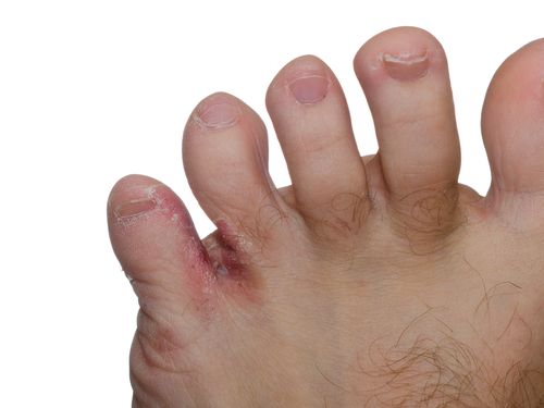
- Presentation may be asymptomatic, but is commonly pruritic, with blisters, scaling and fissuring.
- Tinea Pedis may be classified as
- Interdigital – between the toes and extending onto the plantar surface
- Chronic Hyperkeratotic – moccasin distribution with plantar erythema, scaling, and hyperkeratosis
- Infammatory/vesicular – painful pruritic vesicles with erythema and can be complicated by secondary bacterial infection
- Ulcerative – rapidly spreading erosive lesions with secondary bacterial infection, common in immunocompromised patients.
- More common in men. Unusual in children.
Tests and Imaging
- Microscopy and culture of skin scrapings or de-roofed vesicles.
- Adequate sample size is essential for accurate analysis.
- Swab for secondary bacterial infection
Immediate Treatment
- Application of topical antifungal agents, or combination therapies with hydrocortisone 1%
- Oral terbinafine, itraconazole, or fluconazole may be indicated for severe cases
- Use of antiperspirant may help with hyperhidrosis
- Cease use of occlusive footwear
Possible Referral
- Podiatry for expert debridement and advice on disinfection of footwear and hosiery
- Podiatry for management of hyperkeratosis and hyperhidrosis.
- Dermatologist for inflammatory or ulcerative cases.
![]()